Fluorescence microscopy is a pearl in modern scientific research. It can not only be used to observe fluorescent specimens, but also to conduct routine biological observations. This article will discuss in depth the working principle, advantages of fluorescence microscopy, and its differences and commonalities with ordinary biological microscopes.

How it works:
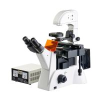
Fluorescence microscopy works based on the phenomenon of fluorescence. It uses short-wavelength light (usually ultraviolet light) to illuminate a sample that has been labeled with fluorescent dyes. These dyes absorb ultraviolet light and fluoresce at long wavelengths when excited. This fluorescent signal is amplified by the microscope's optical system and viewed through the eyepiece.
The advantages of fluorescence microscopy are obvious:
High magnification: Fluorescence microscopy provides high magnification, allowing researchers to observe the details of tiny structures and organelles.
Low cell stimulation: The use of fluorescence microscopy does not cause significant damage to cells, so it is suitable for in vivo staining.
Multiple staining: Researchers can use multiple fluorescent dyes at the same time to make different structures or molecules fluoresce in different colors in the same sample, thereby achieving multiple staining.
Application field:
Fluorescence microscopy has a wide range of applications in scientific research and medical diagnosis, including but not limited to:
Cell biology
Used to observe and study the structure, function and activity of cells.
Molecular biology
Used to detect and quantify specific molecules such as proteins, nucleic acids, etc.
Drug Development
Used to screen for the effects of potential drugs, as well as the distribution and effects of drugs within cells.
Medical diagnosis
Used to detect disease markers and pathological tissue to help doctors diagnose diseases.
Differences and commonalities:
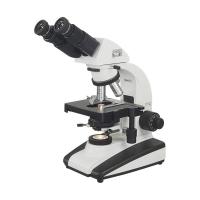
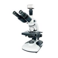
The main difference between fluorescence microscopes compared to ordinary biological microscopes is the use of light sources and fluorescent dyes. Ordinary biological microscopes use visible light sources to observe samples, while fluorescence microscopes use ultraviolet light sources to excite fluorescent dyes, producing a fluorescent signal.
Despite these differences, the two have something in common. They are both microscopes used to observe biological samples that can reveal the mysteries of life. In addition, fluorescence microscopes can also perform general biological observations, so they have a wider range of applications.
In conclusion, fluorescence microscopy is an important tool for modern scientific research, which enables scientists to deeply study the microscopic world, observe and study various structures and processes in cells, molecules and biological samples. Its advantages of multiple staining and high magnification provide strong support for scientific research and medical diagnosis, and will continue to promote progress in the field of science. Fluorescence microscopes, like a light that illuminates the mysteries of microscopic life, reveal endless secrets in the microbial world for us.