I. Introduction
As a precision optical instrument, the microscope has irreplaceable importance in scientific research, education, and industry. It reveals the mysteries of the microscopic world by magnifying tiny objects and enabling people to observe details that are invisible to the naked eye. Microscopy plays a key role, from cell and tissue observation in biomedical research, to microstructure analysis in materials science, to quality inspection and control in industrial production. In addition, with the continuous progress of science and technology, the application of microscopes in frontier fields such as nanotechnology and new materials research has become more and more extensive, which has promoted the development and innovation of science and technology.
The basic functions of a microscope mainly include magnifying microscopic samples, providing clear imaging, and performing high-precision analysis of samples. By utilizing optical lenses or electron beams, microscopes are able to magnify tiny samples to tens or even millions of times to reveal the structure and details of the sample. Microscopes can also incorporate a variety of additional functions such as imaging, shooting, measurement, and analysis, making them a versatile scientific tool. These basic functions make the microscope an indispensable piece of equipment for researchers, engineers, and technicians in their respective fields, greatly improving the efficiency and quality of research and production.

2. Classification of microscopes
1. Optical microscope
Optical microscopes are traditional types of microscopes that utilize visible light and lens systems for imaging. According to the magnification classification, it can be divided into low magnification, popular, high magnification, and ultra-high magnification microscopes. The magnification of the low-magnification microscope is less than 200x, and it has the characteristics of lightweight, small and simple structure, which is suitable for observing animal and plant specimens, plant protection and family education. The magnification of the universal microscope is between 200 and 1000 times, and the structure is straight, simple, and suitable for general biological experiments and inspection and breeding work in agriculture, forestry and animal husbandry. High-power microscopes have magnifications ranging from 1000x to 1600x, and usually use oblique eyepiece structures with higher precision, which are widely used in scientific research and clinical trials. The magnification of ultra-high power microscope can reach more than 10,000 times, and even 1 million times magnification can be achieved by electron microscope, which is mainly used for high-precision scientific research, such as the analysis of viruses and molecular structures.
Use visible light for observation
Classified by magnification
Low-magnification microscopy
Magnification:200 times less
Peculiarity:Lightweight, compact and simple in structure
apply: Observation of animal and plant specimens, plant protection, family education, etc
Popularization microscope
Magnification:200x to 1000x
Peculiarity:Straight and simple structure, suitable for general biological experiments
Disposition:Common eyepiece and objective lens combinations
Apply:Biological experiments, agriculture, forestry and animal husbandry inspection, breeding, etc
High magnification microscopy
Magnification:1000x to 1600x
Peculiarity:Oblique eyepiece structure with high precision
Disposition:High-power eyepieces and objective lenses with a variety of accessories
Apply:Scientific research, clinical trials
Ultra-high magnification microscope
Magnification:More than 10,000 times, electron microscope up to 1 million times
Peculiarity:For ultra-high-resolution observations
Apply:High-precision scientific research

2. Non-optical microscope
Non-optical microscopes use invisible light, such as electron rays, to observe, breaking through the resolution limitations of traditional optical microscopes. Scanning electron microscopy (SEM) provides high-resolution images of the sample surface by scanning the surface of a sample with an electron beam and is suitable for materials science, biology, and nanotechnology. Transmission electron microscopy (TEM) is widely used for detailed analysis of materials and biological structures by passing electron beams through very thin samples to form images, which can achieve atomic-level resolution observation. Scanning probe microscopy (SPM) uses probes to scan the surface surface of a sample, which can analyze the surface structure of a sample at the nanoscale, which is suitable for the research and development of nanomaterials. Due to its ultra-high resolution and wide range of applications, non-optical microscopes have become an indispensable and important tool in modern scientific research.
Observations are made using invisible light, such as electron rays
Scanning Electron Microscopy (SEM)
Principle:Scanning the electron beam to form an image
Apply:Materials Science, Biology, Nanotechnology
Transmission Electron Microscopy (TEM)
Principle:The electron beam passes through the sample to form an image
Apply:Atomic-level resolution observation, structural analysis
Scanning Probe Microscopy (SPM)
Principle:The probe scans the surface of the sample
Apply:Nanoscale surface structure analysis

3. The main components of the microscope
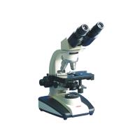
1. an eyepiece
The eyepiece is an important part of the microscope and is responsible for magnifying the objective image and transmitting it to the observer. The eyepiece usually consists of two lenses, and the common magnifications are 5X, 10X, 16X, etc., and the user can choose the appropriate eyepiece according to his needs. The eyepiece not only magnifies the image, but also provides a wider field of view, making observation more comfortable. Some advanced eyepieces are also equipped with a micrometer for precise measurements.
Function:Enlarge the image
2. Objective lens
An objective lens is an optical element in a microscope that is directly imaged, usually mounted at the lower end of the microscope, and provides initial magnification of the sample. Depending on the magnification, objective lenses can be divided into low magnification (e.g., 4X to 20X), high magnification (e.g., 40X to 100X), and oil lens (e.g., 90X to 100X). The choice of objective lens directly affects the magnification and resolution of the microscope, and is one of the key factors affecting the effect of microscopic observation.
Function:Direct imaging
3. Light source
There are two types of light sources in microscopes: transmitted light sources and reflected light sources. The transmitted light source is used for the bottom illumination of the sample, which is suitable for the observation of transparent or translucent samples; Reflective light sources are used to illuminate from above or from the side of the sample and are suitable for opaque samples. The quality of the light source directly affects the brightness, contrast, and clarity of the microscope image, so the selection and adjustment of the light source is crucial to the use of the microscope.
Type:Transmitted light source, reflected light source
4. a focusing device
The focusing device is a component in the microscope that is used to precisely adjust the distance between the sample and the objective lens, ensuring a clear image of the sample. The focusing device is divided into two types: coarse adjustment and fine adjustment, coarse adjustment is used to quickly move the lens barrel to make the sample roughly imaged; Fine adjustment is used to fine-tune the focal length to improve the clarity of the image. The precision and stability of the focusing device are particularly important for the use of high-magnification microscopes.
Function:Precisely adjust the distance between the sample and the objective
5. a stage
A stage is a platform in a microscope that is used to hold and move a sample, usually with a specimen clip and mover. It can be mechanically or electrically controlled, allowing the sample to move precisely in the X and Y directions. The stability and precision of the stage have a direct impact on the efficiency and accuracy of microscopic observations, especially when performing high-magnification observations or microscopic measurements.
Function:Fix and move the sample

Fourth, the operation and maintenance of the microscope
1. Steps
(1) Sample preparation
The sample is properly prepared and fixed on a glass slide, ensuring that the thickness of the sample is uniform and free of bubbles to ensure clarity of observation.
(2) Select the appropriate magnification
Depending on the characteristics of the sample and the observation needs, the observation starts at low magnification first, and then gradually adjusts to a higher magnification to accurately locate and observe the sample in detail.
(3) Adjust the light source
Adjust the intensity and angle of the light source according to the transparency and observation requirements of the sample to ensure uniform illumination of the sample for better image contrast and clarity.
(4) Adjust the focal length
The distance between the objective lens and the sample is adjusted by the coarse and fine adjustment devices to achieve the sample imagemoreExcellent clarity. Coarse adjustments are used for a wide range of adjustments, while fine adjustments are used for fine focusing.
2. Maintenance
The maintenance of the microscope is a critical step in ensuring its long-term stable performance. Regularly clean and disinfect all parts of the microscope, especially the eyepieces, objectives, and stage. These parts come into easy contact with the sample and the operator's hands and need to be gently wiped with a professional cleaning solution and a soft cloth to prevent the accumulation of dust, dirt and fingerprints, thus maintaining the clarity of the optical system.
Microscopes need to be calibrated and checked regularly to ensure the accuracy and reliability of their optical systems and mechanical components. Calibration typically includes alignment of the eyepiece and objective, adjustment of focal length, and examination of measurement function. These steps can help to detect and repair errors or malfunctions that may occur during the use of the microscope in time, ensuring the accuracy of the observations.
To prevent damage to the optics, the operator should avoid direct contact with the lens with his or her hands, and use a dust cover or store it in a dedicated protective case when the microscope is not in use. In addition, the microscope should avoid exposure to high temperatures, high humidity, or strong light to prevent aging and performance degradation of the optical components. Proper care and precautions can effectively extend the life of the microscope.

5. The application of microscopes in different fields
Microscopy plays an important role in biology, especially in cell observation and tissue analysis. Researchers use microscopy to observe cell morphology, division processes, and organelle structure in detail, which is critical to understanding the fundamentals of biology and disease mechanisms. At the same time, microscopy is also used to analyze tissue sections to identify different tissue types and pathological changes, providing a powerful tool for life science research.
Microscopy is also used to study the microstructure and defects of materials. With high-resolution microscopy, scientists can observe the crystal structure, phase interfaces, and microscopic defects of materials, which are critical for material performance analysis, quality control, and the development of new materials. The application of microscopy technology has greatly advanced the development of materials science.
In medicine, microscopes are widely used for the observation and clinical diagnosis of tissue sections. Pathologists use microscopy to analyze tissue samples from patients to identify disease features, such as the morphology and distribution of cancer cells. This precise microscopic analysis is essential for the early diagnosis of diseases and the development of treatment plans, making microscopy an important tool in medical diagnosis.
In addition to this, microscopy is a central tool for studying nanomaterials and surface structures. Scanning electron microscopy (SEM) and scanning probe microscopy (SPM) allow scientists to observe and analyze the surface topography, particle distribution, and chemical composition of materials at the nanoscale. These techniques are important for understanding the properties of nanomaterials and developing new nanotechnology applications.

Through a comprehensive discussion of the classification, structure, operation and maintenance of microscopes, as well as their applications in different fields, we gain an in-depth understanding of the critical role of microscopes as an indispensable tool for scientific research and industrial applications. Microscopy technology has not only helped scientists explore the unknown in the microscopic world, but has also driven significant advances in fields such as biology, materials science, medicine, and nanotechnology.