1. Classification of Microscopes
Scientifically, microscopes are divided into two categories: optical microscopes that work under visible light conditions and non-optical microscopes that work under invisible light conditions (including electron rays). In practical work, people often name microscopes according to their performance and use, and classify them according to their magnification, lighting method, imaging form, mirror body structure, function and use, etc.
(1) Classify by magnification
①Low magnification microscope: the total magnification is below 200 times. This kind of microscope is light in weight, small in size and simple in structure. It is generally equipped with a 10x or 12.5x sun mirror and a 10x or 16x objective lens. It is suitable for observing animal and plant specimens, such as small insects, seed germs, etc., and can also be used for plant protection, family education, etc.
②Popular microscope: a microscope with a total magnification of more than 200 times and less than 1000 times. This kind of microscope often adopts a straight cylindrical structure, and the precision is higher than that of a lower magnification microscope. Generally equipped with 5x, 10x, 16x eyepieces and 4x, 10x, 40x (or 60x) objective lenses. The general combination is less than 640 times, which is suitable for biological experiments, agriculture, forestry, and animal husbandry for inspection and breeding. This model is inexpensive and has a wide range of applications.
③High power microscope: the total magnification is about 1000~1600 times. This kind of microscope often adopts oblique eyepiece tube structure, exquisite shape and high precision. It is equipped with 16 times eyepiece and 100 times objective lens at most. It is suitable for medical, agricultural and animal husbandry scientific research units for laboratory tests, biological research and clinical trials. Sometimes it is equipped with various accessories for extended functions.
④Ultra-high power microscope: the total magnification is more than 10,000 times, and some electron microscopes have reached 1 million times. This microscope is used by specialized research departments to study viruses, molecular structures of substances, analyze crystals, etc.
(2) Classify according to the mirror body structure
① Straight tube microscope: Generally low power, popular type and high power type with low operation requirements all adopt this structure, which is characterized by economical benefits, convenient operation and portability. Its shortcoming is that the observer is relatively hard. If the lens barrel is tilted, the liquid substance on the slice will flow, which will limit the observed object to a certain extent.
②Monocular inclined tube microscope: This kind of microscope adds a prism in the eyepiece tube, so that the optical path is inclined at 45° from the vertical line, and the observer feels relaxed when using it, without changing the horizontal position of the slice. Since natural light is generally used for lighting, the scope of use is limited to a certain extent.
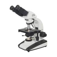
③ Binocular oblique tube microscope: This kind of microscope adds a group of light prisms behind the refracting prism, and uses two eyepiece tubes to observe at the same time. It is suitable for observers to work for a long time, and the eyes will not feel tired. Generally, high-power microscopes and scientific research microscopes adopt this structure, and the distance between the binocular tubes can be adjusted according to the needs of the observer. The base of this kind of microscope is generally made into a box shape, and the interior is equipped with artificial light source lighting, which can provide various lighting requirements, and provides conditions for the microscope to expand functions and expand the scope of use. It is suitable for experiments, scientific research and other purposes.
④ Stereo Microscope
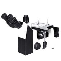
⑤Inverted microscope: The observation object of this microscope is placed above the objective lens, and the objective lens is observed from below, that is, directly from the bottom of the petri dish or beaker, through the quartz glass at the bottom of the container, suitable for some special-purpose research. In addition, metallographic microscopes generally adopt this structure. Since the observed object is an opaque object, the inverted microscope should be equipped with a better epi-illumination light source and a camera device.
(3) Classify according to Ming technology
①Bright field microscope: Generally speaking, microscopes that use transmitted light to illuminate sliced specimens belong to bright field microscopes.
② Dark field (dark field) microscope: it is relative to the bright field microscope, it is equipped with a dark field condenser. In dark field illumination, the light illuminates the observed object along a specific angle direction without entering the microscope objective lens. The light that enters the objective is either diffusely reflected or diffracted by the specimen. In this way, the field of view seen by the observer is dark, and the bright specimen image is set off against the background of the dark field.
③Fluorescence microscope: It is a microscope that uses ultraviolet light to illuminate, and it requires a special light source and filter device that can provide ultraviolet light. When used, the ultraviolet light does not directly enter the observer's eyes, but excites the fluorescent substance in the specimen or the specimen stained by fluorescent infiltration. The observation effect is similar to that of a dark-field microscope, which is to show a bright fluorescent image of the specimen in a dark background. For biological science research.
④Infrared microscope: Infrared light has a greater penetrating ability, and different substances have different degrees of absorption of infrared light. A microscope that makes use of this characteristic to observe objects under infrared light is called an infrared microscope. It is equipped with an infrared light source, which can observe certain opaque objects through transmission, such as infrared light with a wavelength greater than 1.12X10⁻⁶m, which can penetrate single crystal silicon. Infrared light can also be used for episcopic observation of substances with different absorption degrees, and it is a microscope used for scientific research.
(4) Classify according to the way the image is formed
①Phase contrast (phase contrast) microscope: It is often used in biology and medicine. It is a special microscope for observing completely transparent specimens that are inconvenient to be stained. It adds a phase-contrast halo to the microscopic light path, uses the slight difference in refractive index between the specimen and the encapsulation medium around the slice to produce an optical path difference, and produces light interference imaging for observation. It is suitable for observing the growth, movement, proliferation and fine structure of living cells in living conditions. Therefore, it is a necessary tool for modern biological research such as microbiology, cell engineering, and hybridoma technology.
②Polarizing microscope: The main difference between a polarizing microscope and an ordinary microscope is that the microscope is equipped with two polarizing elements. That is, polarizer and analyzer. The polarizer is placed under the condenser, and the analyzer is placed above the objective. The stage is made circular according to the needs of observation and can be rotated, with a dial on it to measure the rotation angle of the inspected crystal. Polarizing microscope uses polarized light to decompose into two paths of refracted light in the crystal, which interfere with each other and form images. It is generally used for the detection and identification of biological liquid crystals and inorganic salt crystals.
③Interference microscope and optical section microscope: Both are microscopes for optical metrology using the principle of interference imaging after light is reflected on two similar interfaces. It is mainly used to measure the roughness of the surface of the object, and it is used as an inspection and identification instrument in industry.
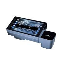
④Projection microscope and TV display microscope: Both are formed by adding a projection device or a TV camera device to an ordinary microscope, which can send out the microscopic image and display it on the screen. This kind of microscope requires a relatively stable machine, a large field of view, high precision, and a relatively strong lighting source. It is suitable for more people to observe, teach and demonstrate at the same time.
(5) Classification by functional use
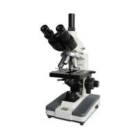
①Teaching microscope: Generally, it is modified and expanded from a monocular inclined tube microscope. Its eyepiece tube is divided into two or more branches by prism, which can allow more than two persons to observe the same specimen at the same time. When the main observer adjusts and observes the desired image, other observers also observe the image at the same time and experience the whole adjustment process. It is very suitable for the instructor to guide the students and carry out physical teaching.
②Dissecting microscope: It is used for observation by doctors during microsurgery, also called operating microscope. The fuselage can be adjusted according to the operation situation, and it is also suitable for several people to observe at the same time. The teaching dissection microscope has simple structure and low magnification, and is used for biological anatomy demonstration.
③ Analytical microscope: sometimes called research microscope, is a high-end microscope for scientific research. This kind of microscope has relatively complete functions, and can observe under conditions such as bright field, dark field, phase contrast, polarized light, and fluorescence under transmitted light illumination. At the same time, it also has a variety of accessories, which can be used for projection, photography, measurement and automatic analysis and recording. It is a multifunctional model, suitable for colleges and research institutes to engage in various experimental analysis.
④Metallographic microscope: It is a microscope dedicated to observing and identifying opaque objects such as metal sections, minerals, materials, and crystals. This microscope cannot use a transmitted light source and needs to illuminate the specimen with epi-light. In order to obtain images, the microscope is generally equipped with a photographic light splitter and a photographic eyepiece. Due to the large field of view, the metallographic microscope needs to be equipped with a plan achromatic objective lens and a wide-angle compensation eyepiece, so that the field of view of the imaging surface of the obtained micrograph can be large and clear.
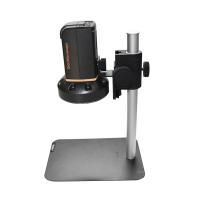
⑤Endoscope: It is a special-purpose testing instrument that has been improved and developed on the basis of medical endoscopes in recent years. It is equipped with an objective lens group on the top of the probe tube of the endoscope, and a peep window which can change the observation angle is installed in front of the objective lens group, and an observation eyepiece which can be finely adjusted is installed on the other end of the probe tube. Insert the probe tube into the sealed inner cavity of the object to be observed, and turn on the supporting light source to observe the microscopic endoscopic image. At present, endoscopic microscopes are not only used in clinical medicine, but also used in industry as flaw detection of airtight cavities inside machines. In particular, such endoscopic microscopes have been commonly used for routine safety inspections of combustion chambers within engines of jumbo jet aircraft. When used with a 135 single-lens reflex camera, it can also record the slight scars inside the machine for timely treatment and avoid major losses.
(6) Introduction to non-optical microscopy
Non-optical microscopes are a new member of the microscope family. With the development and progress of science and technology, in order to improve the magnification and resolution of the microscope, non-optical microscopes have made great progress, mainly including the following varieties.
①Ultrasonic microscope: It is made by applying ultrasonic technology to a microscope. Utilizing the characteristics that ultrasonic waves can penetrate some objects that light waves cannot penetrate, ultrasonic microscopes can be used to observe the internal structure of some opaque objects, such as the structure under the opaque conductive film of integrated circuits, untreated red blood cells, etc. It is a widely used scientific instrument.
② Electron microscope: Electron microscope is a new type of microscope that uses the principle of magnetic lens to focus cathode rays to image and observe microscopic particles. Its magnification can reach more than 1 million times, and its resolution can reach 2X10-''m tiny structures. It is a professional instrument used in high-tech fields.
③Scanning electron microscope: The general electron microscope adopts the transmission method and can only observe extremely thin samples. A scanning electron microscope uses electron beams to directly scan and observe the surface of the object to be observed. In this way, not only the sample preparation is simple, but also the uneven fracture surface can be directly observed, the magnification is high, the focal depth is long, and the imaging has a three-dimensional effect. The scanning electron microscope also has the ability to analyze the chemical composition of the surface of the observed object. It is a professional high-tech equipment and is widely used in biology, physics, medicine, and electronic technology.
④X-ray microscope: It is an instrument that analyzes and observes the X-ray spectrum of the observed substance to determine the structure and composition of the substance. Applicable to physical observation and analysis of matter.
⑤Scanning tunneling electron microscope: It is a new type of electron scanning microscope successfully developed in the 1980s. It is generally suitable for new material research and microscopic surface test analysis.
With the development of science and technology, the variety of microscopes is constantly expanding. The professional microscope structure is the comprehensive application of three sciences and technologies: light, mechanics and electricity. The types of microscopes that have been widely used at present can be summarized as follows, see Table 2-31.