Preliminary research on the application of illuminance meter in the measurement of the light transmission of eye tissues. Direct ophthalmoscope is used as the light source, and the sclera transillumination method is used to measure the illuminance reflected by the pupil area with the illuminance meter, so as to evaluate the light transmission of the main light-transmitting tissues in the eye. light performance. In addition, the light-transmitting properties of isolated cornea, lens and other eye tissues were tested in a similar way. Discuss the clinical application of this group of detection methods.
Objective: To detect the light-transmitting properties of living and isolated ocular tissues and explore the possibility of the application and clinical application of this detection method.
Eye tissues have different degrees of light transmission, such as: cornea, aqueous humor, lens, vitreous body, etc., have high light transmission. Although the choroid and sclera act as a darkroom, they are not completely opaque. How permeable are these tissues to light? Can it be measured accurately? Can it be measured in vivo? This experiment focuses on the preliminary research and discussion of the above problems.
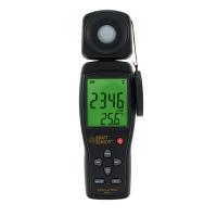
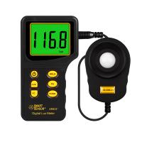
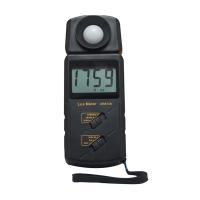
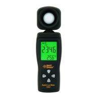
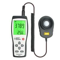
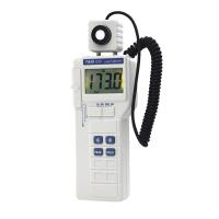
Equipment: illuminance meter (accurate to 0.1LUX); direct ophthalmoscope, used as light source.
Method: In a dark room, use a direct ophthalmoscope as a light source, and transmit the light source at the outer canthus and sclera of living rabbits. Note that the light source should be closely attached to the sclera to prevent light leakage. At the same time, the illuminance meter was placed in the pupil area of the rabbit's eye, and the distance between the illuminance meter and the cornea was shortened as much as possible, and the consistency of the distance should be ensured for each measurement. In this way, the light transmission of the eyes of living rabbits can be measured. After recording the experimental data, the cornea, vitreous body, choroid and sclera of the rabbit eye were removed respectively, and the light transmittance was measured respectively between the illuminance meter and the light source. This allows the light transmission of isolated ocular tissue to be measured.
Discussion: According to the experimental results, it can be seen that the choroid and sclera cover most of the light, so that only a very weak illuminance reading can be obtained in the pupil area when measuring in vivo. If the intensity of the light source and the concentration of the light source are further increased, the light that can enter the eye through the sclera-choroid layer and reflect from the pupil will inevitably increase. Since the illuminance meter is accurate to 0.1, increasing the light intensity can greatly reduce the experimental results. mistakes. In addition, it is also important to maintain a stable and consistent brightness of the light source.
We believe that it is entirely feasible to measure the luminous flux of eye tissue more accurately in vitro or in vivo, and this (especially in vivo measurement) will be of great help to clinical work. For example, it can be applied to the grading of cataract opacity, aqueous humor turbidity, corneal edema or corneal clouding, macula and leukoplakia, etc. It enables objectification of many indicators that were previously graded only by subjective observations. We will continue to explore and strive to make this method more widely used in clinical work.