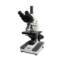
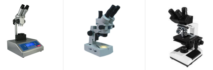
The two binoculars of a binocular microscope make viewing and analyzing laboratory specimens more effortless than a monocular microscope. Read on to better understand the parts and functions of binocular microscopes.
eyepiece
Binocular binoculars contain the optics of the microscope and provide the user with secondary magnification of the "objective" or object being viewed (usually a specimen on a glass slide).
mechanical platform
A mechanical stage holds an object or slide sample under an objective lens for viewing and allows the sample to move left, right, forward and backward for examination.
objective lens and objective lens
The objective turret contains several rotating objectives, usually three, that magnify the image of objects on the stage below.
spotlight
A built-in light in the base provides illumination for the viewing area. Light from the lamp passes through the condenser, which focuses the condenser on the viewing area of the microscope.
Microscope Tubes and Prisms
The microscope barrel supports the binoculars of the binoculars and the multirefringent prism - where the light rays are split and directed towards the binoculars.
Scientists use many tools to expand their view of things they cannot see, either because they are too small to see with the eye or because they are too far away. Some instruments help scientists observe other objects, including your body. Some tools magnify objects, while others penetrate tissue, water, or inorganic materials to reveal what lies beneath the surface.
microscope
Microscopes use light or electrons to magnify small objects such as microorganisms. A standard laboratory microscope, often called a compound microscope because it has two lenses to magnify an object, uses light for magnification. The objective lens closest to the object being magnified works together with the optical lens closest to the eye. Compound microscopes can magnify up to 2,000 times. Electron microscopes, on the other hand, can magnify up to 500,000 times, but cannot magnify living objects because objects need to be observed in a vacuum. Scientists use two types of electron microscopes: transmission electron microscopes and scanning electron microscopes, with transmission electron microscopes being the more common of the two.
telescope
Scientists use telescopes to observe distant stars, planets and galaxies. Telescopes can magnify distant objects and can also use light to magnify objects. However, telescopes need to collect a lot of light. For this, the telescope needs a large objective lens. A telescope's ability to gather light is more important than its ability to magnify. When using a telescope, you change the focal length of the optics, not the objective lens, which is your eye. With a microscope, you adjust the objective lens instead of the optical lens.
X-ray
Although you may primarily think of x-rays as a way to examine bones in the body, x-rays have been used outside of orthopedic clinics. Scientists use X-rays not only for medical purposes but also to visualize solid structures buried underground. Airports use X-rays to scan luggage and people for potentially harmful solid objects. X-rays send electrons through objects until they hit a solid object. The electrons collide with atoms in the target object, generating energy visible in X-rays. A computerized tomography scan, or CT scan, combined with X-ray images to create a 3-D image of an organ or structure can help detect tumors and other soft tissue and organ abnormalities.
ultrasound
Scientists use ultrasound machines to outline soft tissue in the body by bouncing sound waves off the tissue. A computer forms an image from the sound waves. One of the common uses of ultrasound is pregnancy. According to Dr. Stephen Carr of Brown University, 70 percent of women in the United States have at least one fetal ultrasound. Underwater sonar, short for sound navigation and ranging, is used by fishermen to detect fish and find boats and structures underwater.
Magnetic resonance imaging
Magnetic resonance imaging (MRI), better known, uses magnets and radio waves to create detailed slices of organs and tissues, which are then put together to create images. These machines can detect tumors and abnormalities in soft tissues and organs.