如果没有在现实世界中探索和放大的能力,发现和发明的进展将受到损害。用于研究和探索生命科学的显微镜有很多类型。According to目的,放大倍数,样品类型和应用,应选择合适的 显微镜。下面列出了一些常用的显微镜。
简单显微镜
它被认为是第一台显微镜。它是由列文虎克在创建17 个世纪。 他将凸透镜和镜架结合在一起。它可以将特殊的物种放大 200到300倍(更像是放大镜)。显微镜非常简单,但是功能强大,因为它能够提供有关生物标本的信息。
钟表匠使用一台简单的显微镜放大并观看手表的小部分。珠宝商,棕榈商,皮肤专业人士也使用它。它用于放大书中字母的图像,纤维的质地,布的线以及图章和雕刻的细节。如今,由于引入了带有第二透镜的其他显微镜,简单的显微镜已不再使用,从而提高了图像质量。
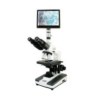
复合显微镜
在复合显微镜中,载玻片借助于放置在下方的灯泡 进行照明。 样品被2枚透镜放大,其中2枚放置在载玻片附近,称为物镜,而另一枚放置 在顶部附近,称为目镜。 它们提供了一个二维图像,可以According to镜片的强度进行调整.
复合显微镜的设计各不相同,但通常是相当标准的,这使任何人都可以轻松使用它们。复合微型显微镜的优势在于,它们对于学生,业余爱好者以及科学家而言都是负担得起的,并且可以放大到 更高的倍率。一个主要的缺点是它们具有较低的分辨率,因此,像更高级的显微镜一样,图像永远不会清晰。
荧光显微镜
英国科学家乔治·斯托克斯(George G. Stokes)于1852年第一次描述了荧光。当他观察到矿物氟石被紫外线激发时发出红光时,他创造了荧光一词。他指出,荧光发射的波长比激发光的波长长。 荧光显微镜是一种特殊的工具,因为它可以观察目标分子发出的荧光。这样做是由于向目标细胞中添加了特定的荧光试剂,因此当激发光施加到目标细胞时它们会发出荧光。
Chlorophyll, vitamin A, collagen, riboflavin, etc. can be seen under this microscope without the use of fluorescent pigments because of their autofluorescent properties. Other substances such as cellulose, glycogen, protein, starch and Y chromosome also need to be stained with fluorescent dyes. By using fluorescent dyes, we can see specific proteins in cells.
confocal microscope
In a confocal microscope, visible light comes from a laser source, which makes it different from a stereo and compound microscope. The microscope has a series of scanning mirrors through which the laser scans the sample, and the images are then assembled into a computer for display on a screen. There are no eyepieces in this microscope. They provide a two-dimensional image that can be adjusted according to the strength of the lens.
In contrast to fluorescence microscopy, this microscope uses point illumination and a pinhole in an optically conjugate plane in front of the Detector to eliminate out-of-focus signals, hence the term "confocal". Since only light generated by fluorescence very close to the focal plane can be detected, the optical resolution of the image is much better than that of a widefield microscope. However, since much of the light from the sample's fluorescence is blocked at the pinhole, this improvement in resolution comes at the expense of reduced signal intensity, thus requiring long exposures.
scanning probe microscope
Scanning probe microscopy is used to study surfaces at the nanoscale. T by probing his image forms. The probe scans the surface of the sample and collects data by touching it. The collected data is displayed as a computer image.
The scanning tunneling microscope (STM) was the first scanning probe microscope. This is the first approach for a technology with atomic resolution capabilities. STM uses an electric current as the scanning tip, which is a limitation because only conducting or semiconducting materials can be studied under this microscope. Thanks to the development of SPM, scientists and engineers are able to see structures and details at a finer resolution without the need for rigorous sample preparation. Developments in expertise and technological advancements have greatly expanded the capabilities of SPM. These microscopes are scanned in ordinary air rather than a vacuum, as is the case with electron microscopes.
electron microscope
In an electron microscope, a beam of electrons is used instead of light to illuminate an object, thus magnifying to the Angstrom level. There are many variations of electron microscopy such as scanning (SEM), transmission (TEM) and scanning transmission (STEM).
Sample preparation in electron microscopy is also intense, on which the image quality depends to a large extent. Depending on the properties of the sample, a combination of dehydration, freeze fixation, embedding, staining, conductive coating, etc. steps may be required. The microscope can be used in applications such as cryobiology, protein localization, electron tomography, cell tomography, cryo-electron microscopy, toxicology, bioproduction and viral load monitoring, particle analysis, pharmaceutical quality control, 3D tissue imaging, and virology.
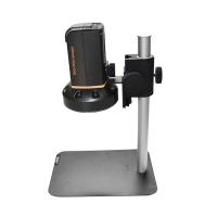
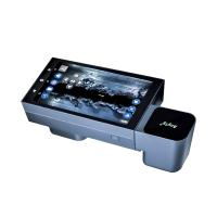
digital microscope
Digital Microscope It was invented in Japan in 1986. It uses computers to see objects invisible to the naked eye. They exist in two forms, with or without eyepieces. With a USB cable, it can be connected with a computer monitor. With the help of computer software, it can be displayed on a computer screen as a magnified sample. Movies can be recorded, while still images can be saved in computer memory. Saved images can be saved for a longer period of time by e-mail. It can be used by researchers, students, hobbyists and makers.
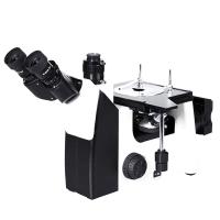
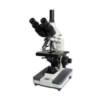
stereo microscope
Stereo Microscope A stereo microscope differs from a compound microscope mainly in that it has two eyepieces instead of one. Stereo microscopes produce three-dimensional images when two eyepieces send different images to the left and right eye. Specimens are typically illuminated from above rather than below, making stereo microscopes ideal for dissection, fabrication, inspection, circuit board work, or use with any opaque specimen. T hey is cheap, which makes it ideal for professionals, hobbyists, and everyone else in the industry. Magnification is lower since individual cells cannot be seen.
pocket microscope
It's small, but impressively capable. It's durable and portable, so it's easy for a scientist or student to use. It is used for handheld imaging of various objects outdoors or in the laboratory. Magnification varies from 25x to 100x. They use batteries and LED lights.