microscope terminology
Achromatic depiction of the area observed by a standard microscope. With an achromatic microscope, the edges of the viewing area are curved. This curve changes the perception of samples at the edges of a circular viewing area and makes them appear rounder and longer. This is similar to objects seen at the edge of a magnifying glass.
Achromatic Plan: A depiction of the viewing area of a microscope in which distortions at the edges of the viewing area are compensated for by a more expensive lens system; a true representation of the entire viewing area can be seen.
Pole Microscope: A microscope in which the focus adjustment knob moves the pole on the entire top of the unit, rather than on the arm. This type is not as robust as traditional arm microscopes.
Refractive Index: The relative speed of light passing through a material.
Resolution or Resolution: The ability to distinguish fine details using a magnifying glass instrument. Also known as the ability to distinguish two points at a specified distance.
Total Magnification : The total magnification of the sample, obtained by multiplying the magnification of the eyepiece by the magnification of the objective.
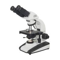
microscope parts
The arm area on the back of the microscope that supports the eyepieces and objectives. Handles and parts of handles.
The bottom of the fundamental range. If there is a light source, it is usually placed. The extended rear of the base also serves as a handle for lifting and carrying the microscope.
Route focus knob: used to adjust the position of the objective lens. This should be adjusted while continuing to ensure that the objective does not hit the slide. Stop as soon as you see the object through the eyepiece. Any further adjustment of the knob could break the slider or lens on the objective, which is very expensive to replace.
Fine focus knob: used to focus the specimen to a desirable position after observing the specimen through the route focus knob. Adjust very slowly to prevent contact between the objective and the sample.
Illuminator: The light source for the microscope.
Objective or Numerical Aperture: This part of the compound microscope is the lens closest to the sample.
Ophthalmic Lens: In a compound optical microscope, this is the lens closest to the viewer.
Oil Immersion Lens: This is the 100x objective lens. The lens is small for high magnification and high resolution. Due to its small size, it is important to let as much light as possible pass through the lens. By immersing the lens in oil, the refraction (bending) of light is eliminated, since glass and oil have approximately the same refractive index. In this way light is maximized and gives the sharpest images. If you use an oil immersion lens without oil, the image will be blurry and have poor resolution.