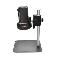
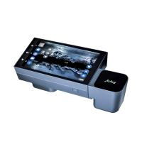
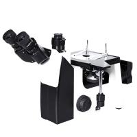
Different Types of Microscopes
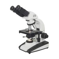
Compound Microscope: There are two sets of lenses (objective and eyepieces) that use visible light as the source of illumination.
Darkfield microscope: has a device that scatters the light from the illuminator, making the specimen appear white against a black background.
Electron Microscope: Uses a stream of electrons instead of light to produce images. They can enhance images of viruses, ribosomes, proteins, lipids and even small molecules.
Fluorescence microscopy: uses an ultraviolet light source to illuminate specimens that will fluoresce. Typically, fluorescent dyes or antibodies have been added to the specimen being viewed.
Phase Contrast Microscopy: Allows the use of special condenser lenses to examine structures inside cells. They take advantage of different refractive indices and allow the examination of living organisms, since it is not necessary to stain the cells to provide good component differentiation. The final image is a combination of light and dark.
Frequently Asked Questions About Microscopes
Q: Why do I need immersion oil with the 100x objective?
A: In order to achieve powerful magnification, the lens needs to be cut very small. Therefore, it is important to allow as much light as possible to pass through the lens so that the observer can see the object in question. The oil changes the refraction of the light so the light passes through the lens instead of being bent and scattered in all directions.
Q: Which is better: plan achromat or achromat?
A: Both microscopes are well suited for viewing specimens in the center of the field of view. However, if it is necessary to count bacteria anywhere in the field of view or to distinguish the shape of bacteria, a plan achromatic microscope is better suited. These microscopes feature a lens system that compensates for distortion at the edges of the field of view so that specimens appear exceptionally sharp.
Q: How do I know the total magnification of what I'm viewing?
A: The first thing to determine is the magnification of the IOL; that is the lens closest to the viewer. The magnification of this lens will be etched into the metal. The standard magnification of the eyepiece is 10x. Then you need to find the magnification of the objective lens. That is the lens closest to the sample. This number will also be inscribed into the goal. These two magnifications are then multiplied together to obtain the total magnification. For example, if you have a standard 10x eyepiece, and you use a 40x objective, the total magnification will be 400x.