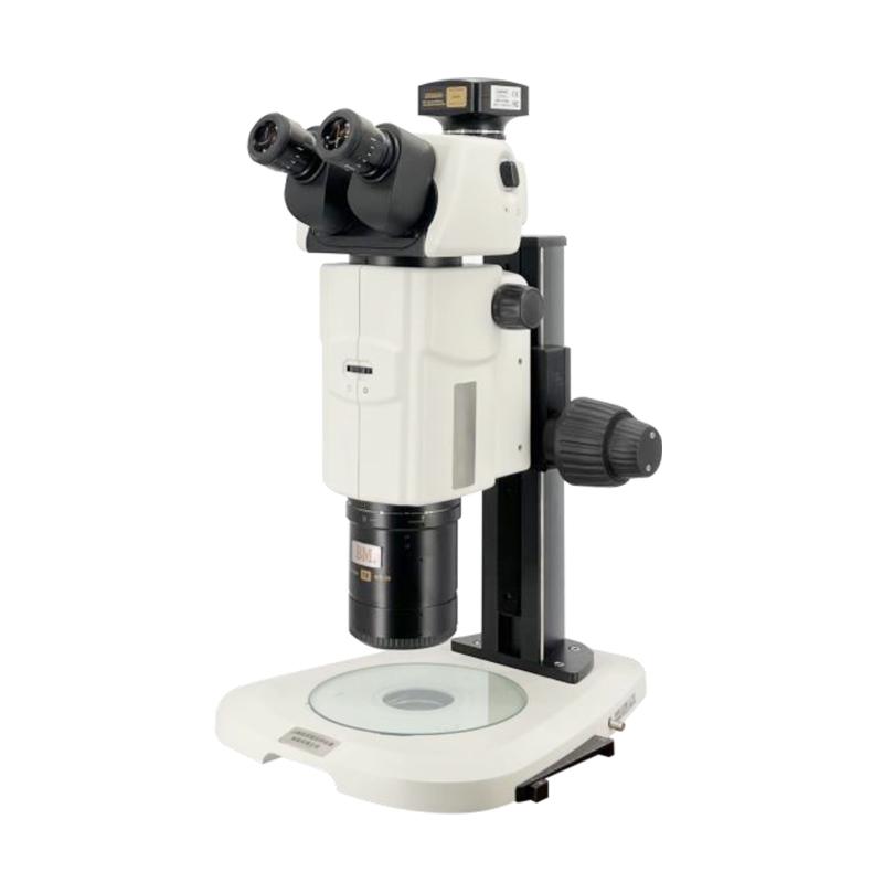

VARNISING XTL-BM-18TD Computer Parallel Light Body Microscope with 3D Stereoscopic Viewing
Built-in OCC oblique light illumination, enhanced uneven sample contrast ratio, effective illumination area diameter of 60mm, eyepiece field of view with three-dimensional stereoscopic observation and super depth of field expansion.
 SHSGBM
 XTL-BM-18TD


Introduction
The XTL-BM-18TD computerized paralleluristic stereo microscope is commonly used for high-contrast observation and imaging of protein crystallization processes and crystals, as molecular biology, cell biology, neurobiology, developmental biology, embryology, and systems biologyMacroscopic to microscopic high-resolution observation and imaging research tools for zebrafish, mice, nematodes and other model organisms and various transparent samples, microscopic cell tissues, and subcellular structures of brightfield and relief contrast; Can be upgraded to a fluorescence observation and imaging system.
Features
Built-in OCC oblique illumination enhances the contrast of uneven samples, and the effective illumination area is 60mm in diameter;
High-resolution 50OLP/mm apochromatic 1X objective;
Built-in aperture diaphragm;
Three-dimensional observation and ultra-depth depth of field expansion under the eyepiece field of view;
Strong scalability, to achieve fluorescence, dark field, polarized light and other observations.
Standard configuration
| High focal point adjustable focus eyepiece | Standard configuration | |
| Working distance 60mm, Wavelength range: 380-700nm | ||
| Total magnification | The field of view of the object | |
| 10X/23 | 7.5X | 30.7mm |
| 135X | 1.7mm | |
SHSGBM XTL-BM-18TD Computer Parallel Light Body MicroscopeSpecifications
| LIST | VALUE |
|---|---|
| magnification | 7.5x-135x |
| Zoom Ratio | 1:18 |
| Magnification Range | 7.5x-135x |
| Focusing device | Manual focusing device with built-in CounterWeight and focusing mechanism Shaft thick, fine-tuning knob (60 + 90mm) |
| Binocular pupil distance adjustment range | 52-76mm |
| diopter compensation | Vision correction range + 5~ -5 |
| Color temperature compensation | Color temperature automatic compensation |
| Light source | LED Cold Light source; Built-in OCC; Illuminator: OCC oblique light illumination, illumination area: diameter 60mm; built-in aperture diaphragm |
| Computer imagery system | (1) Digital CMOS camera, 12 million pixels. With geometric measurement analysis software, the software for point, line, circle and arc, straightness, roundness, area and other measurements; (2) MCL-Z adapter mirror; (3) computer self-purchased |
SHSGBM XTL-BM-18TD Computer Parallel Light Body Microscope Packing list
host X1, manual X1, certificate X1, warranty card X1
[Note] Because the manufacturer's packaging may be updated or upgraded, the detailed packaging list shall be subject to the latest standard configuration of the manufacturer.
FAQ
- Working Principle and Application Analysis of Confocal Microscope
- VITINY F300 Series Portable Storage Digital Microscope Operating Instructions
- Overview of the different types of microscopes
- Characteristics of Electron Microscopes and Differences Between Several Microscopes
- What are the defects of optical microscopes? Industry common sense
- 37XB InvertedMicroscope Instruction Manual
- How does a digital microscope work?
- VISION MV-BR600 High Definition Industrial Inspection Microscope Operation Instructions
- Microscope section measurement of coated Film thickness
- VISION MV-BR600 High Definition Industrial Inspection Microscope Operation Instructions