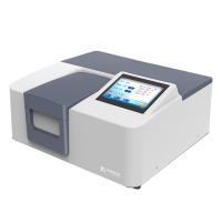
A Spectrophotometer is a scientific instrument that breaks down light with its complex composition into spectral lines. The measurement range generally includes the visible light region with a wavelength range of 380nm to 780nm and the ultraviolet region with a wavelength range of 200nm to 380nm. Different light sources have their own unique emission spectra, so different illuminants can be used as light sources for the instrument.
Today we will study the composition, working principle and characteristics of the Spectrophotometer . We hope that after reading this article, you will have a general understanding of Spectrophotometer s.
The basic principle
Absorption spectra of substances are measured using ultraviolet light, visible light, infrared light, and laser light. The method of qualitative and quantitative analysis of substances and material structure analysis using this absorption spectrum is called spectrophotometry. The Spectrophotometer has high sensitivity, fast measurement speed and wide application range. Among them, UV/Vis spectrophotometry is one of the basic methods needed for biochemical research.

Spectrum: Light is an electromagnetic wave that can be represented by a wavelength "λ". The electromagnetic spectrum consists of a series of continuous wavelengths with different properties. For biochemistry, the important wavelength regions are visible light and ultraviolet light.
The wavelength of light is the distance between two adjacent wave crests.
The propagation of light is composed of mutually perpendicular electric field component "E" and magnetic field component "H".
λ=C/ν (λ represents the wavelength. C represents the speed of light. V represents the frequency)
The number of waves passing through a fixed point per unit time.
The ultraviolet region can be divided into ultraviolet (near ultraviolet) and vacuum ultraviolet (far ultraviolet). Since absorption cells (also called sample cells, cuvettes, etc.) and optics and oxygen absorb light at wavelengths below 190 nm, conventional UV measurements focus on the near-UV region, ie, 200 nm to 400 nm. The visible region is 400nm to 800nm.
The molecules that make up matter are in a certain state and are in constant motion. Molecular motion can be divided into translation, rotation, vibration, and the motion of electrons in the molecule. Each state of motion is at a certain energy level, so the energy of a molecule is the sum of all its energy levels.
Each energy of a molecule has a series of energy levels. Energy levels are not arbitrary, but have quantifiable characteristics. Normally, the molecule is in the ground state. When it absorbs a certain amount of energy and transitions to an excited state, an absorption spectrum is produced.
Molecular rotation, vibration, and electronic energy level transitions generate rotational, vibrational, and electronic spectra, respectively.
According to the principle of quantum mechanics, the energy state of molecules changes in a jumping manner according to certain rules. Under the irradiation of incident light, when a molecule absorbs light, its energy increases discontinuously. The frequency and energy difference of the two energy levels need to satisfy the following relationship:
E=E2- E1=h
E1 and E2 represent the energy of the initial energy state and the final energy state, respectively. The greater the energy difference between the initial energy state and the final energy state, the higher the frequency (that is, the shorter the wavelength) of absorbed light. The lower the frequency of the light (ie the longer the wavelength).
Since the absorption is discontinuous, a series of dark bands of absorption appear in certain parts of the light. Due to the large difference in the energy of molecular rotation, vibration and electronic energy level transition, their absorption spectra appear in different spectral regions. The molecular rotation energy level difference is very small, △E<0.05 electron volts (ev), and the absorption of the molecular rotation spectrum appears in the far infrared or microwave region.
The vertical difference of vibrational energy levels is large, E=0.05~1.0 eV, and the vibrational spectrum appears in the mid-infrared region. The electronic energy level difference is large, E=1~20ev, so the spectrum obtained by electronic transition appears in the spectral region of visible light, ultraviolet light or shorter wavelength.
Calculation principle
Spectrophotometers use light sources that can produce multiple wavelengths. And through a series of spectroscopic devices, a light source with a specific wavelength is generated. After light passes through the sample to be measured, part of the light is absorbed. Calculate the absorbance of the sample to convert to the concentration of the sample. The absorbance of a sample is directly proportional to the concentration of the sample.
When monochromatic light radiation passes through the solution of the measured substance, the amount absorbed by the substance is proportional to the concentration of the substance and the thickness of the liquid layer (optical path length). The relationship is as follows:
A=-lg(I/I0)=-lgT=kLc
In the formula: A is the absorbance.
I0 is the incident monochromatic light intensity.
I is the transmitted monochromatic light intensity.
T is the light transmittance of the material.
K is the molar absorptivity.
L is the optical path of the substance to be analyzed, that is, the side length of the cuvette.
C is the concentration of the species.
The selective absorption wavelength of light by a substance and the corresponding absorption coefficient are physical constants of the substance. When the absorption coefficient of the pure substance under certain conditions is known, the test substance can be prepared into a solution under the same conditions, and its absorbance can be measured, and the content formula of the substance in the test substance can be calculated from the above formula.
In the visible light region, except for some light-absorbing substances, many substances do not absorb light themselves, but it can be determined by adding a color developer or processing the color under certain conditions. Therefore, it is also called colorimetric analysis.
Since there are many factors affecting the color depth during color development, and instruments with poor monochromatic light purity are often used, standard substances or reference substances should be used at the same time.